A newborn swallows its first meals a few drops at a time: thick, yellow milk that many nurses call “liquid gold.” This first milk is not sterile. In 2016, Alba Boix-Amorós, Maria Carmen Collado and Alex Mira counted what it carries: about a million bacterial cells in each milliliter, some twenty drops.
The count held as later milk came in. At about 800 milliliters a day, a little over three cups, a baby takes in ten million to a hundred million bacterial cells. Older counts, which grew the bacteria on lab dishes, found about a hundred times fewer. Those cells are an early share of what NPR called an invisible universe (Figure 1).

Figure 1. A tour of the microbes on and in a body. Drawn by Ben Arthur for NPR (2013). The video repeats an old estimate of ten microbial cells for every human cell; a 2016 recount by Ron Sender, Shai Fuchs and Ron Milo put it closer to one to one. Video and thumbnail: NPR, Standard YouTube License.
Which bacteria end up in a gut is not left to chance. In 1899, Henri Tissier, a children’s doctor at the Pasteur Institute in Paris, looked at babies’ stool under a microscope. In breastfed babies, one bacterium crowded out almost all the rest. Its rods often split at one end into a Y, so he named it bifidus, Latin for “split in two.” Today it is called Bifidobacterium. In bottle-fed babies, and in babies sick with diarrhea, it was far scarcer, and a mixed crowd took its place. The milk seemed to pick the winner.
Part of the answer is food. Human milk also carries a family of sugars the baby cannot digest. Most of them reach the colon, the last stretch of the gut, untouched. In 2006, Robert Ward, Bruce German and colleagues at the University of California, Davis, purified those sugars. They gave them to bacteria as their only food. One common gut bacterium could not live on them. Bifidobacterium infantis, one of Tissier’s Y-shaped bacteria, could. A year later, the team tested five kinds of Bifidobacterium, and infantis grew three times as dense as the rest. Every feed has two eaters: the baby, and the baby’s bacteria.
I have fed babies both ways. My first daughter had only breast milk. My second weighed more than 10 pounds at birth and was hungry before my milk arrived, so I breastfed her and we topped her up with formula. Her sister’s diapers had been mustard yellow. Hers were light brown, and they differed in texture, smell and how often they came. Tissier would have put both diapers under his microscope and counted the Y-shaped rods.
Bacteria settle wherever a body meets the outside world: in the gut, on the skin, in the mouth and nose, in the vagina. Each place grows its own crowd. Count every crowd, with the fungi and viruses beside them, and the whole is called the human microbiome.
“The microbiome is a term used to describe the specific collection of microorganisms (such as fungi, bacteria and viruses) that exist in a particular environment.”
Say you stand in the dairy aisle this week with a probiotic yogurt in one hand and a plain tub in the other. The probiotic costs twice as much. Its label promises billions of live bacteria for your gut, and the foil lid is cold under your thumb. Into the cart it goes, this week and most weeks after, fifty-two tubs a year. The label counts what goes in. It says nothing about the crowd already living there to meet it.
Milk feeds a baby’s bacteria at every meal, and a grocery aisle sells adults more. If bacteria keep arriving from the first swallow to the fifty-second tub, what are they to the body? Systems thinking answers a question like that by asking what else each part is connected to. In a gut, each kind of bacterium is fed, held back or crowded out by the others and by the body it lives in.
In 2024, Ziqi Ma and three colleagues, at Heidelberg University Hospital and Sun Yat-sen University, drew on more than 700 papers to answer that question. Their review sets out eight ideas for how microbes and a body fit together. The last four, on how the microbes work for health and why a body needs them, are part two of this series. This piece follows the first four. Biology now counts microbes the way it counts genes, all at once, and a count names the parts without the ties between them. The review opens on a map of where the microbes live.
Where the Crowds Live
Ma’s first figure pins a percentage to each place on a drawn human body where bacteria live. The gut gets 29 percent and the mouth 26, close behind. The skin, the airways and the urogenital tract share the rest. The review calls the gut the most densely populated of the five.
I traced those numbers to a pie chart the Human Microbiome Project published in 2009, then tried to replicate it. The project’s website listed every bacterium it had sequenced or queued for sequencing, with the body site each came from. The Internet Archive saved two earlier copies of that list, from March and April 2009. I counted both by the pie’s own rule: one strain, one count, sorted by body site. A strain is one line within a species, the way a beagle is one breed of dog.
Neither copy matched the pie, and the two did not match each other. In March, the gut held 58 percent of the list. By April it held 42, and in the pie, 29 (Figure 2). The list grew, and the shares moved with it. The shares measure what the project chose to sequence. When a probiotic label or a headline next quotes a share of the microbiome, the question to ask is what was counted.
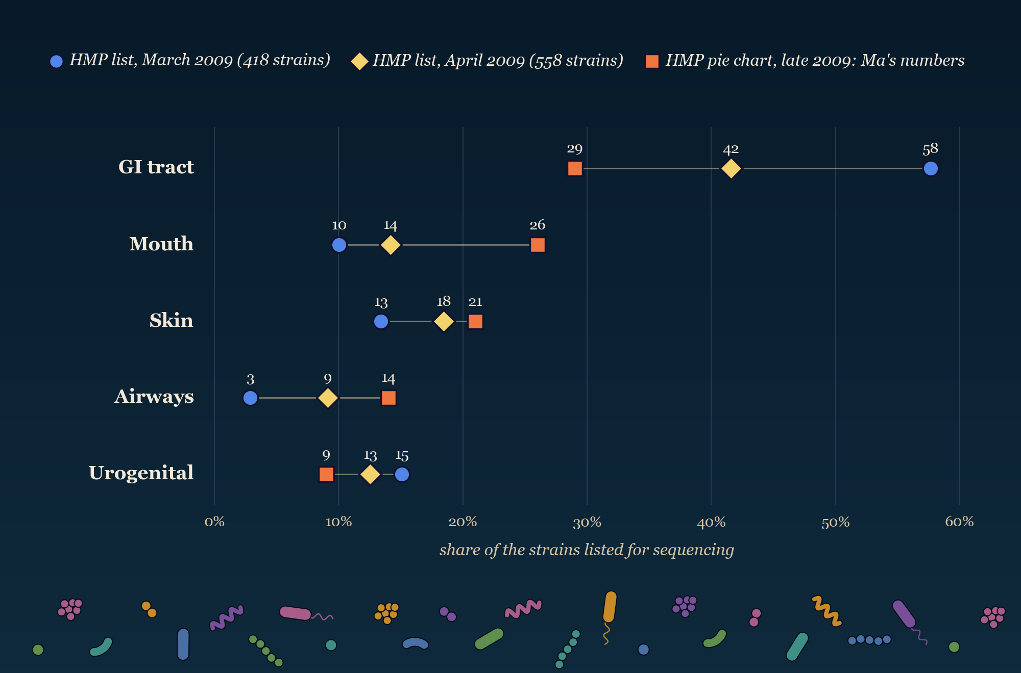
Figure 2. The five percentages in Ma et al.’s 2024 review (squares), beside my recount of two earlier versions of the same list (circles and diamonds). Each marker is a share of the strains the Human Microbiome Project (HMP) had sequenced or queued for sequencing from that place, not a share of the bacteria living there. Squares from NIH HMP Working Group (2009), Fig. 3, as reprinted in Ma et al. (2024), Fig. 1; circles and diamonds counted for this piece from the project’s online strain lists, as saved by the Internet Archive. Chart drawn for this piece.
Where bacteria live takes a different count. Ma’s five shares look close to even. By cell count, they are far from it. Most of a body’s bacteria live in the colon. A single gram of stool, about the weight of a paper clip, holds some 90 billion bacteria, eleven times the number of people on Earth. The plainest test of what so many do is a birth with none waiting.
What Goes Missing
Doctors in Houston placed David Vetter inside a plastic bubble within seconds of his birth, in September 1971. He had severe combined immunodeficiency, an inherited disease that leaves the body almost no immune defense. His older brother had died of it as a baby.
Everything that went into the bubble was sterilized first: food, water, clothes, toys. Filtered air flowed in without stopping. His parents touched him through rubber gloves set into the plastic wall. In 1983, doctors gave him bone marrow from his sister, hoping it would build him an immune system. The marrow carried a virus no test had found. He died of a cancer it caused in February 1984, at twelve.
David is the closest thing to a germ-free person in the medical record, and he says little about what germs do for a body. His immune system had failed from birth, so any difference in him could come from the disease. For a cleaner answer, scientists turned to animals.
At the Lobund Laboratory at the University of Notre Dame, rats were delivered by cesarean section into sealed steel tanks and raised there on sterilized food, generation after generation. In 1963, Helmut Gordon, Bernard Wostmann and Edith Bruckner-Kardoss compared ten germ-free rats from the colony with twelve ordinary rats of the same stock. The two groups weighed the same. The germ-free hearts pumped about 30 percent less blood per minute for their weight, and not one germ-free rat reached the ordinary range. In a second set of rats, the germ-free ones also carried about a fifth less blood.
Later studies, gathered in Ma’s review, added to the list. Germ-free animals have a swollen cecum, the pouch where the small intestine meets the large one, about twice its usual size in rats. They have a smaller thymus, the gland that trains immune cells, and fewer immune cells in the gut wall.
The changes reach the brain. A tight seal of cells, the blood-brain barrier, keeps most of what travels in the blood out of the brain. In 2014, Viorica Braniste, Sven Pettersson and colleagues at the Karolinska Institute in Stockholm injected a blue dye into the blood of mice, three to a group. In ordinary mice, the dye stayed inside the blood vessels. In germ-free mice, it leaked into the brain itself. The leak starts before birth and lasts into adulthood. When the team gave adult germ-free mice the gut bacteria of ordinary mice, the seal tightened. Butyrate alone, a fatty acid that gut bacteria make from fiber, tightened it as far as in ordinary mice.
Ma’s team gathers these findings under one name: germ-free syndrome. Taking the microbes away does not leave a clean body. It leaves a body with missing parts. “A gut devoid of microorganisms,” they write, “cannot be considered a complete intestine.”
Germ-free syndrome is Ackoff’s definition of a system, run as an experiment. Each part can change how the whole behaves, but never alone. The mice lost the microbes in their gut, and the seal around their brains changed.
Anyone who has finished a course of antibiotics has run a small, short version of this experiment. In a Danish study, twelve healthy men took a mix of strong antibiotics for four days. Their gut bacteria took about six weeks to come back close to where they had been. Six months later, nine common species were still gone from most of the men.
In the weeks after a course, the question is what changed besides the infection it treated: digestion, appetite, a rash. The person to send this section to is a friend halfway through a course of pills. Whatever the missing microbes did, the rats and mice could not do it with their own genes.
Whose Genes Count
On a rosebush, an aphid that lacks the genes to feed itself presses its mouthparts into a stem and drinks. Sap is mostly sugar water. It holds too few of the amino acids an animal needs to build proteins, and the aphid cannot make them.
Bacteria called Buchnera live inside special cells in the aphid’s body and make the missing amino acids for it. The bacteria have lost so many of their own genes that they cannot live anywhere else, and the aphid cannot live without them. A mother aphid passes them straight into her eggs. Almost every aphid starts life with its bacteria in place.
People start differently. By most evidence, a human baby grows in the womb without live microbes. The first ones arrive at birth, from the mother’s skin, gut and birth canal, then from breast milk, from the father and from everyone who holds the baby. They keep arriving for life. By about age three, a child’s gut community settles into something close to an adult’s.
The bet on how many genes a person carries ended near 20,000. The bacteria in one gut carry far more. In 2010, the European MetaHIT team sequenced stool from 124 people. They counted 3.3 million distinct microbial genes, about 150 times the human count. Any two people differ in only about 0.1 percent of their own DNA, Ma’s team notes. The microbes may explain more of what sets one person apart. How much more, no one knows yet.
Ma and colleagues split a person’s genes into two sets. The innate genome is the one built from egg and sperm. It is inherited by fixed rules, and it does not change after birth. The adaptive genome is the set of genes the microbes carry. It is picked up over a lifetime. It changes with what a person eats, the drugs they take, where they live and who they live with. The innate genome changes over many generations. The adaptive genome can change in days (Figure 3).
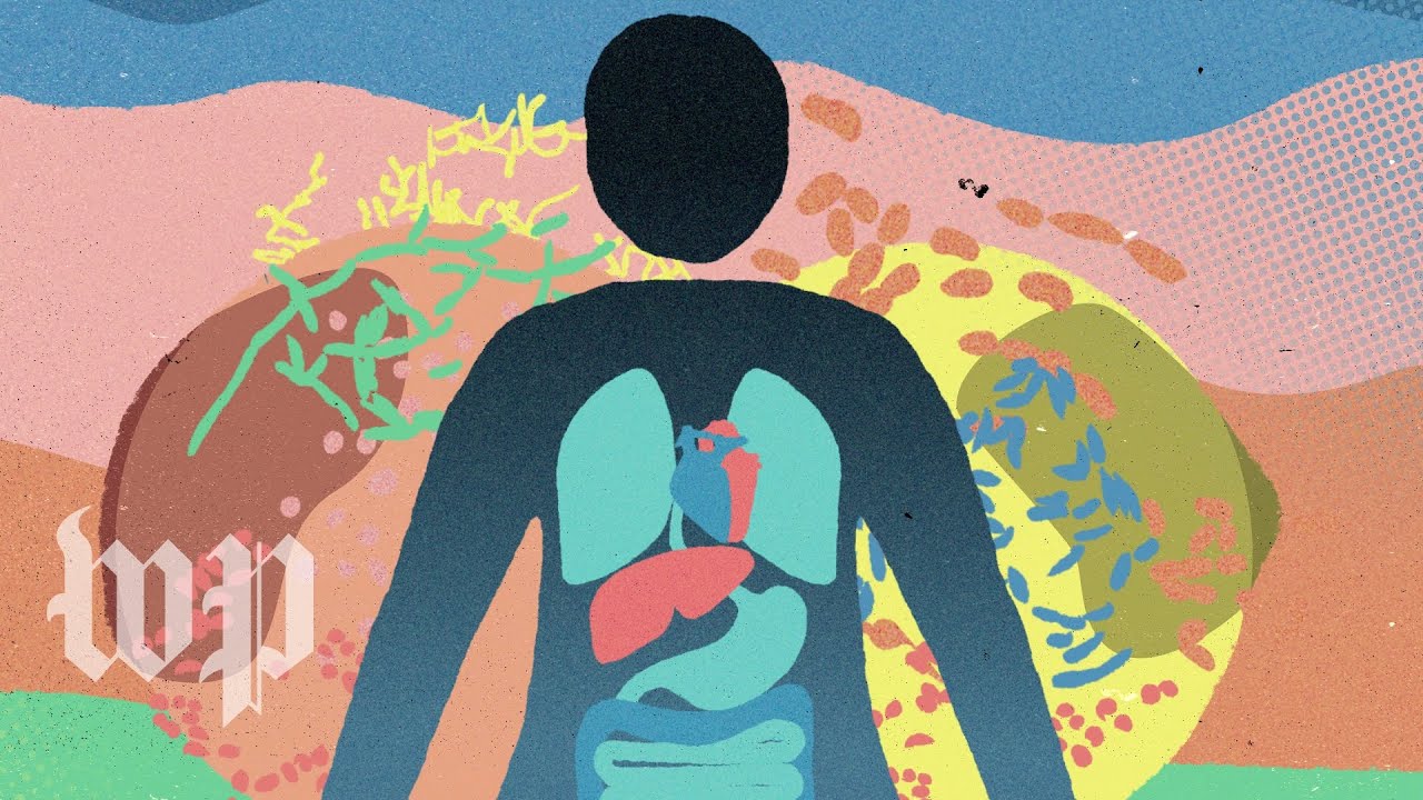
Figure 3. About 20,000 human genes against millions of microbial ones, and why food can change the second set but not the first, from The Washington Post (2021), with food and mental health reporter Mary Beth Albright. The mouse and fly studies it cites are early work. Video and thumbnail: The Washington Post, Standard YouTube License.
Living together is enough to share some of those genes. In 2023, Mireia Valles-Colomer and colleagues traced bacterial strains across more than 9,700 samples from people around the world. People in one home shared about 12 percent of their gut strains and 32 percent of their mouth strains. The longer they had lived together, the more mouth strains they shared. The samples show who shares strains. They cannot show who passed them to whom.
A new baby, a new partner or a new roommate starts that sharing under one roof. The question for a household is who brought what in. When a stomach bug moves through a home and skips one person, the germ was the same. The hosts were not.
Who the Host Is
In South Africa, Christina Gosmann and Douglas Kwon’s team tracked one germ, HIV, in 236 young women. They tested the women again and again. None had it at the start, and all were 18 to 23. Thirty-one became infected during the study.
The team had also sampled the bacteria in each woman’s vagina. Some women had a mixed community, with many kinds of bacteria and few Lactobacillus. Others had a community dominated by one species, Lactobacillus crispatus. The mixed group was more than four times as likely to become infected. Not one of the women dominated by L. crispatus became infected. The mixed communities came with more of the activated immune cells that HIV infects.
A study that follows people cannot settle what caused what. The women with mixed communities may have differed in other ways too, and the bacteria could be a marker as much as a cause. Animals can be tested more directly. In 1955, B. P. Phillips and colleagues placed Entamoeba histolytica, an amoeba that causes dysentery, into the guts of germ-free guinea pigs. The amoeba did no harm. Feed the guinea pigs one kind of common gut bacterium first, and the amoeba damaged the gut wall.
A cleaner test was run on a mosquito. Aedes aegypti carries dengue, a virus that brings fever, crushing joint pain and, in the worst cases, bleeding. Many insects carry a bacterium called Wolbachia inside their cells, but this mosquito normally does not. Scott O’Neill’s team at Monash University moved a strain of Wolbachia from fruit flies into Aedes aegypti. Inside the mosquitoes that carried it, the virus grew poorly. Like the aphid’s bacteria, Wolbachia passes from mother to eggs, so it can spread through a wild population on its own.
From 2017 to 2020, Adi Utarini and colleagues at Gadjah Mada University tested those mosquitoes in Yogyakarta, Indonesia. They split a part of the city, home to about 313,000 people, into 24 areas and released Wolbachia mosquitoes in half of them, chosen at random. Volunteers left buckets of water and mosquito eggs around the neighborhoods, and the eggs hatched into mosquitoes carrying the bacterium. In the areas that got them, dengue fell by 77 percent, and hospital stays for dengue by 86 percent. It was one trial, in one city. The mosquitoes were the same species, and the virus was the same virus. The bacteria living inside the mosquito changed how often it passed the virus on.
Whether a germ makes someone sick depends on the germ, on the person’s own genes and on the microbes already there. Ma calls the person and their microbes together a meta-host. Doctors have long asked why one germ makes one patient very sick and leaves another well, and why one transplant takes while another fails. Ma’s team points to the meta-host as part of the answer. The one person a stomach bug skipped at home was a different meta-host from the rest of the household. The crowd that person carried is one of three places to look for why. The host had always been a crowd, and part of that crowd lives in every human mouth.
What the Body Keeps in Check
On 17 September 1683, Antonie van Leeuwenhoek wrote to the Royal Society in London about his teeth. He was a cloth merchant in Delft who ground his own lenses. He scraped the white paste from between his teeth, thick as wet flour, and looked at it through one of his lenses. It was alive.
Some of the tiny animals darted through it “as a Jack or Pike does thro the water.” Others “spun about like a Top.” He kept his teeth “usually very clean,” he wrote, and still they were there. He looked at the teeth of two old men, one who lived soberly and one who drank brandy and wine every day, and found them there too.
Three centuries later, scientists still argue about what to call them. In 1992, the immunologist Velio Bocci, at the University of Siena in Italy, called the gut’s bacteria “the neglected organ,” and the label spread. In 2022, Alessandro Fucarino, Francesco Cappello and colleagues at the University of Palermo answered that the label does not fit. An organ is built from tissues that grow from the embryo, and microbes arrive from outside. In a 2026 essay, the microbial ecologist Allan Konopka points out that the right picture depends on the level someone looks from.
In their 2024 review, Ma’s team looks from the level of tissues. Next to the four kinds in biology textbooks, epithelial (the body’s linings), connective, muscle and nervous, they propose a fifth. They call it a “slave tissue,” a term borrowed from sociology, to say the body is in charge. This piece calls it a managed tissue.
The managing never stops, because bacteria left alone keep multiplying. More cells make more cells, a reinforcing loop that slows only when the food runs out. So the body sets limits of its own, and one of them can be seen under a microscope.
In 2008, Malin Johansson, Gunnar Hansson and colleagues at the University of Gothenburg, in Sweden, looked at the lining of the colon in five mice. It wore two coats of mucus, together about as thick as two human hairs. The outer coat was loose and full of bacteria. The inner coat, packed tight and stuck to the wall, held none. In mice bred without the main mucus protein, bacteria lay against the cells of the wall, and the colon became inflamed.
Mucus is one limit among several. Antibodies released into the gut keep microbes in their places. Proteins in the gut and in milk lock away iron and zinc that bacteria need to grow. Leeuwenhoek’s tooth cleaning in Delft belongs on that list. So do the milk sugars from the opening. Only some bacteria can eat them, so a mother’s milk helps choose which ones grow in her baby’s gut, as Tissier saw in Paris. The microbes help the body only while the body keeps them in check.
The same managing reaches the dinner plate. Onions, apples and whole-grain bread carry fiber, food the body cannot digest and many gut bacteria can, much as the milk sugars feed Bifidobacterium. When fiber runs short, some of those bacteria turn to the mucus coat instead (Figure 4). What goes on the plate tonight helps decide what the colon’s bacteria eat.
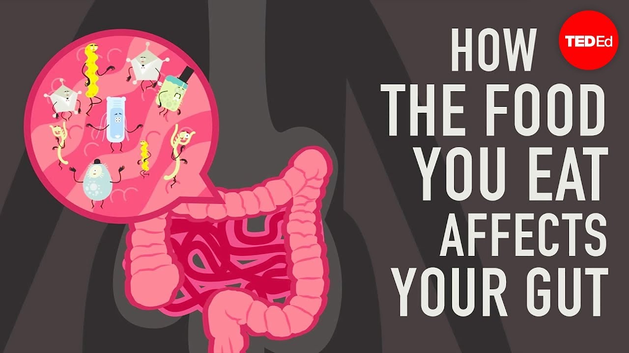
Figure 4. Fiber as food for gut bacteria, and what hungry bacteria do to the mucus lining, from TED-Ed (2017): lesson by Shilpa Ravella, animation by Andrew Foerster. CC BY-NC-ND 4.0.
By the sink, a toothbrush with splayed bristles has been doing the managing twice a day.
A Closing Invitation. The milk in the opening fed two eaters at once. So did every meal and every dose in this piece: whatever reaches a body reaches its microbes too. Three ways to watch for the second eater.
- Map who shares. From where you sit, picture what your home passes around: the toothbrush cup, the shared towel, the pillow, the spoon at dinner. People who live together share about a third of their mouth strains, and more the longer they stay. Who has lived beside you longest, and what crowd do you two now carry in common?
- Trace the last bug. Think back to the last stomach bug that went through your home. Who caught it first, who caught it last, and who was skipped? The same germ had its chance at each of them. What else might the one who stayed well have been carrying?
- Turn the tub over. At the store this week, pick up one probiotic yogurt or capsule box and read the back, the plastic cold in your hand. What does it count: bacteria made, bacteria alive, or bacteria that stay? Which crowd is already in your gut to meet them?
Tonight, somewhere, a baby feeds. Each swallow carries bacteria in, and sugars that only some of them can eat. Both eaters grow at every feed, and a scale weighs only one of them.
Where This Came From
The four ideas here are the first half of Ma’s eight; the order is this piece’s own. Ma’s review traces the lineage. Lynn Margulis called a host with its microbes a holobiont in 1991, and Richard Jefferson later named their combined genes the hologenome. The aphid fits that word well. Ma’s team argues that people do not, because contact is not inheritance, and so they split a person’s genes in two.
Intellectual Honesty Note. This piece describes what milk carries, not how anyone should feed a baby. The two daughters and their feeds are my own. The dairy aisle in the stakes paragraph is hypothetical. The squares in Figure 2 come from a 2009 Human Microbiome Project pie chart that counts bacteria sequenced or queued for sequencing. Ma’s text reads them as density. It also drops the pie’s blood (1 percent) and eye (0 percent) slices, so its five places add up to 99 percent. My recount (lists saved March 6 and April 10, 2009) groups body sites by name: gut and stomach under GI tract, vagina and urine under urogenital. Strains listed with no site, 1 and 4 percent, count in the totals but get no marker. No later copy of the list was saved, so the pie itself could not be recounted. Ma et al. (2024) is a narrative review, and its ideas are proposals, not settled findings. Tissier’s findings are reported from later accounts; his 1900 thesis was not opened. The germ-free results come from rodents and guinea pigs. The mucus result is from mice, and “two human hairs” is this piece’s gloss of its 150 micrometers. The Entamoeba result is reported from summaries of the paper, which was not opened. Leeuwenhoek’s words are from the Royal Society’s English abstract of his letter, printed in 1684. Ma’s team counts eight ideas, though one of their own figures lists seven; part two folds the eighth, cell-microbe co-ecology, into its set-point section.
References
Albright, M. B. (2021, October 30). What is the gut microbiome? [Video]. The Washington Post. YouTube. https://www.youtube.com/watch?v=d-Ln9NNj2KY
Boix-Amorós, A., Collado, M. C., & Mira, A. (2016). Relationship between milk microbiota, bacterial load, macronutrients, and human cells during lactation. Frontiers in Microbiology, 7, 492.
Bocci, V. (1992). The neglected organ: Bacterial flora has a crucial immunostimulatory role. Perspectives in Biology and Medicine, 35(2), 251–260.
Braniste, V., Al-Asmakh, M., Kowal, C., Anuar, F., Abbaspour, A., Tóth, M., et al. (2014). The gut microbiota influences blood-brain barrier permeability in mice. Science Translational Medicine, 6(263), 263ra158.
Fucarino, A., Burgio, S., Paladino, L., Caruso Bavisotto, C., Pitruzzella, A., Bucchieri, F., & Cappello, F. (2022). The microbiota is not an organ: Introducing the muco-microbiotic layer as a novel morphofunctional entity. Anatomia, 1, 186–203.
Gordon, H. A., Wostmann, B. S., & Bruckner-Kardoss, E. (1963). Effects of microbial flora on cardiac output and other elements of blood circulation. Proceedings of the Society for Experimental Biology and Medicine, 114(2), 301–304.
Gosmann, C., Anahtar, M. N., Handley, S. A., Farcasanu, M., Abu-Ali, G., Bowman, B. A., et al. (2017). Lactobacillus-deficient cervicovaginal bacterial communities are associated with increased HIV acquisition in young South African women. Immunity, 46(1), 29–37.
Indriani, C., Tanamas, S. K., Khasanah, U., Ansari, M. R., Rubangi, Tantowijoyo, W., et al. (2023). Impact of randomised wMel Wolbachia deployments on notified dengue cases and insecticide fogging for dengue control in Yogyakarta City. Global Health Action, 16(1), 2166650. https://doi.org/10.1080/16549716.2023.2166650
Johansson, M. E. V., Phillipson, M., Petersson, J., Velcich, A., Holm, L., & Hansson, G. C. (2008). The inner of the two Muc2 mucin-dependent mucus layers in colon is devoid of bacteria. Proceedings of the National Academy of Sciences, 105(39), 15064–15069. https://doi.org/10.1073/pnas.0803124105
Kennedy, K. M., de Goffau, M. C., Perez-Muñoz, M. E., Arrieta, M.-C., Bäckhed, F., Bork, P., et al. (2023). Questioning the fetal microbiome illustrates pitfalls of low-biomass microbial studies. Nature, 613(7945), 639–649.
Konopka, A. (2026, May 6). Systems thinking [Substack post]. https://thinkmicrobe.substack.com/p/systems-thinking
Leeuwenhoek, A. van. (1684). An abstract of a letter from Mr. Anthony Leevvenhoeck at Delft, dated Sep. 17. 1683, containing some microscopical observations, about animals in the scurf of the teeth. Philosophical Transactions of the Royal Society of London, 14(159), 568–574. https://doi.org/10.1098/rstl.1684.0030
Ma, Z., Zuo, T., Frey, N., & Rangrez, A. Y. (2024). A systematic framework for understanding the microbiome in human health and disease: From basic principles to clinical translation. Signal Transduction and Targeted Therapy, 9, 237. https://doi.org/10.1038/s41392-024-01946-6
National Human Genome Research Institute. (n.d.). Microbiome. In Talking Glossary of Genomic and Genetic Terms. https://www.genome.gov/genetics-glossary/Microbiome
NIH HMP Working Group, Peterson, J., Garges, S., Giovanni, M., McInnes, P., Wang, L., et al. (2009). The NIH Human Microbiome Project. Genome Research, 19(12), 2317–2323. https://doi.org/10.1101/gr.096651.109
NPR. (2013, November 5). The invisible universe of the human microbiome [Video], illustrated by B. Arthur. YouTube. https://www.youtube.com/watch?v=5DTrENdWvvM
Palleja, A., Mikkelsen, K. H., Forslund, S. K., Kashani, A., Allin, K. H., Nielsen, T., et al. (2018). Recovery of gut microbiota of healthy adults following antibiotic exposure. Nature Microbiology, 3(11), 1255–1265.
Pertea, M., Shumate, A., Pertea, G., Varabyou, A., Breitwieser, F. P., Chang, Y.-C., et al. (2018). CHESS: A new human gene catalog curated from thousands of large-scale RNA sequencing experiments reveals extensive transcriptional noise. Genome Biology, 19, 208.
Phillips, B. P., Wolfe, P. A., Rees, C. W., Gordon, H. A., Wright, W. H., & Reyniers, J. A. (1955). Studies on the ameba-bacteria relationship in amebiasis: Comparative results of the intracecal inoculation of germfree, monocontaminated, and conventional guinea pigs with Entamoeba histolytica. American Journal of Tropical Medicine and Hygiene, 4(4), 675–692.
Qin, J., Li, R., Raes, J., Arumugam, M., Burgdorf, K. S., Manichanh, C., et al. (2010). A human gut microbial gene catalogue established by metagenomic sequencing. Nature, 464(7285), 59–65.
Ravella, S. (2017, March 23). How the food you eat affects your gut [Video]. TED-Ed. YouTube. https://www.youtube.com/watch?v=1sISguPDlhY
SciShow. (2016, April 11). Aphids: Weird poop, weirder babies [Video]. YouTube. https://www.youtube.com/watch?v=hNzeshvjZdw
Sender, R., Fuchs, S., & Milo, R. (2016). Revised estimates for the number of human and bacteria cells in the body. PLOS Biology, 14(8), e1002533. https://doi.org/10.1371/journal.pbio.1002533
Shigenobu, S., Watanabe, H., Hattori, M., Sakaki, Y., & Ishikawa, H. (2000). Genome sequence of the endocellular bacterial symbiont of aphids Buchnera sp. APS. Nature, 407(6800), 81–86.
Shearer, W. T., Ritz, J., Finegold, M. J., Guerra, I. C., Rosenblatt, H. M., Lewis, D. E., et al. (1985). Epstein-Barr virus-associated B-cell proliferations of diverse clonal origins after bone marrow transplantation in a 12-year-old patient with severe combined immunodeficiency. New England Journal of Medicine, 312(18), 1151–1159.
Tissier, H. (1900). Recherches sur la flore intestinale des nourrissons (état normal et pathologique) [Doctoral thesis]. Faculté de Médecine de Paris.
Utarini, A., Indriani, C., Ahmad, R. A., Tantowijoyo, W., Arguni, E., Ansari, M. R., et al. (2021). Efficacy of Wolbachia-infected mosquito deployments for the control of dengue. New England Journal of Medicine, 384(23), 2177–2186.
Valles-Colomer, M., Blanco-Míguez, A., Manghi, P., Asnicar, F., Dubois, L., Golzato, D., et al. (2023). The person-to-person transmission landscape of the gut and oral microbiomes. Nature, 614(7946), 125–135.
Walker, T., Johnson, P. H., Moreira, L. A., Iturbe-Ormaetxe, I., Frentiu, F. D., McMeniman, C. J., et al. (2011). The wMel Wolbachia strain blocks dengue and invades caged Aedes aegypti populations. Nature, 476(7361), 450–453.
Ward, R. E., Niñonuevo, M., Mills, D. A., Lebrilla, C. B., & German, J. B. (2006). In vitro fermentation of breast milk oligosaccharides by Bifidobacterium infantis and Lactobacillus gasseri. Applied and Environmental Microbiology, 72(6), 4497–4499.
Ward, R. E., Niñonuevo, M., Mills, D. A., Lebrilla, C. B., & German, J. B. (2007). In vitro fermentability of human milk oligosaccharides by several strains of bifidobacteria. Molecular Nutrition & Food Research, 51(11), 1398–1405.
Yatsunenko, T., Rey, F. E., Manary, M. J., Trehan, I., Dominguez-Bello, M. G., Contreras, M., et al. (2012). Human gut microbiome viewed across age and geography. Nature, 486(7402), 222–227.
Zhou, M. (2024, August 1). David Phillip Vetter (1971–1984). Embryo Project Encyclopedia, Arizona State University. https://embryo.asu.edu/pages/david-phillip-vetter-1971-1984