Stanley Lambchop woke up flat. In Jeff Brown’s 1964 book Flat Stanley, a bulletin board fell on him. It pressed him half an inch thick. Stanley slid under doors. His parents mailed him to California (Figure 1). His brother Arthur flew him in the park like a kite. At a museum, he posed as a painting and caught two thieves. When Stanley tired of being flat, Arthur pumped him back to round, the way a bicycle tire fills.
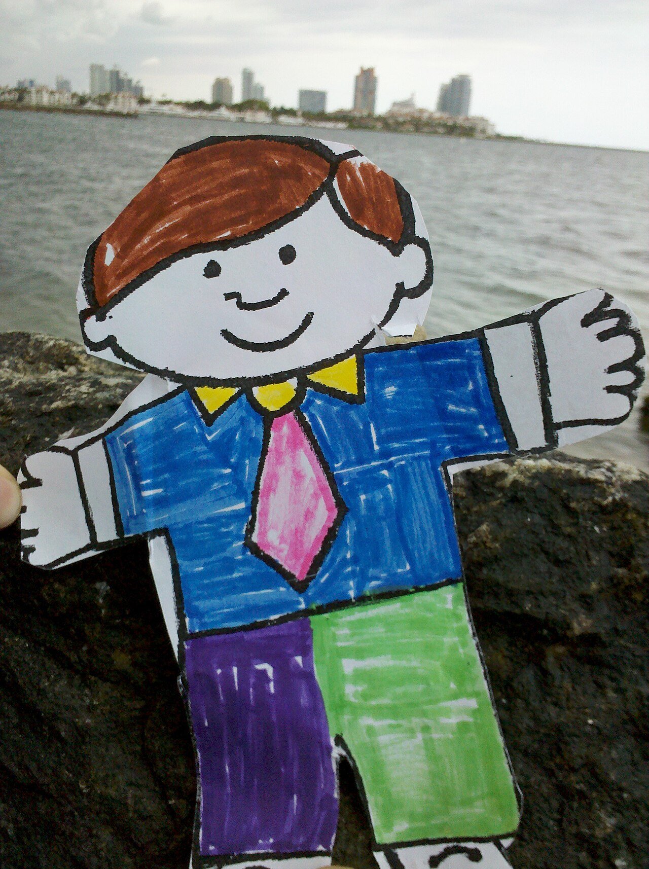
Figure 1. Half an inch thick and still traveling. A paper Stanley from the Flat Stanley Project in Miami Beach, 2011. Photo by Llamabr, CC BY 3.0, via Wikimedia Commons.

A real person pressed flat would keep the crease. Still, the book gets one thing right about proteins, the working parts of a body. The same boy, in a new form, does new jobs: a letter, a kite, a painting. A protein is a chain of small building blocks, and the way the chain folds decides what it does. A body builds its proteins from about 20,000 genes. Some proteins, pulled out straight, fold back on their own, the way Stanley went back to round. Others, once folded wrong, never do, and they can teach the wrong fold to their neighbors.
A day is full of folding. A paper airplane glides across a room because of its creases, and the same sheet folded wrong drops nose first to the floor. A fitted sheet, still warm from the dryer, goes into the closet neatly only with a second pair of hands. A road map folded back in a hurry at a gas station, along the wrong creases, never lies flat in the glovebox again.
In a previous essay, genes were recipes, and copies of them decided which dishes got made. The full spread of dishes, every protein a body can make, has a name: the proteome. A liver cell and a nerve cell keep the same cookbook and serve very different dishes (Figure 2), and the dishes change from hour to hour. The first part of this series followed the letters themselves, a field of sprites in a long row, and the imp that inspects them for slips. The second followed one verse of those letters, a single gene, copied onto a thread and carried out to the counter. The dishes are proteins, and each one is built, then folded, before it can do its job.
“The protein complement of an organism coded for by its genome.”
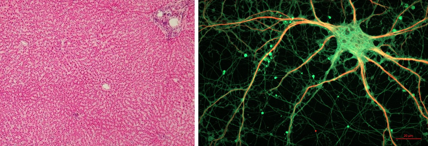
Figure 2. Different cells, different dishes. Left: human liver, its cells packed in rows; liver cells make albumin, a protein that carries cargo through the blood. Right: a rat nerve cell, its long arms built on a form of tubulin found in nerve cells, stained green, with a second protein, MAP2, in red along the branches. Liver: Department of Histology, Jagiellonian University Medical College, CC BY-SA 3.0, via Wikimedia Commons. Nerve cell: ZEISS Microscopy, CC BY 2.0, via Wikimedia Commons.


For twenty years, the clipboard form at a US blood drive, at work or at church, asked one question with nothing to do with germs. You take the folding chair with the pen cold in your hand and the air smelling of alcohol swabs, a paper cup of orange juice waiting on the table. “From 1980 through 1996, did you spend time that adds up to three (3) months or more in the United Kingdom?” A yes pressed you flat, like the bulletin board on Stanley: turned away for life. A patient who needed a transfusion lost a donor. The reason was one wrongly folded protein, carried in British beef. In 2022, the FDA dropped the question. If you once checked yes, the chair at the next drive may be yours again, like Stanley pumped back to round. So how does a fold, with no germ in it, keep a donor out of the chair for two decades?
How the Builders Read
In the second part, a quick imp named Quill copied one verse of the field onto a thread. Quill touches down at a cluster of stockier imps, the builders, round-shouldered and built for handling. It hands the thread over and lifts off again.
The builders read the thread three beads at a stretch, always three. Each triplet acts like one word rather than three letters. A builder reads the first triplet and reaches into a supply shelf stocked with tiny colored droplets. The builder pulls out the one droplet that triplet calls for, clips it onto a growing chain, and moves on to the next three beads (Figure 3).
Figure 3. One short verse read through: the strand Quill reads, the thread Quill makes, and the droplet the builders clip on for each three-bead word. The verse is invented; the matching and the codon readings are real.
If the inspector had missed a slip back on the field, the builders would never know. The wrong letter is copied into the thread like any other. Its triplet may call for a different droplet, and the builders clip it on without a pause. Some of the changed letters on a person’s gene test report began as slips like this one. A letter gained or lost does more harm: every triplet after it is read out of step.
At last a triplet arrives that calls for no droplet at all. The nearest builder reaches for the shelf, comes back empty-handed, and shrugs. The builders let go of the chain: a long, straight row of droplets with no shine to it yet.
Narrator: The builders are ribosomes, and the droplets are amino acids. Each three-bead word is a codon. Four letters can make 64 different codons. Of those, 61 call in one of twenty amino acids. The other three are stop codons, which tell the ribosome to let the chain go. The whole reading, thread to chain, is translation.
“The process through which information encoded in messenger RNA (mRNA) directs the addition of amino acids during protein synthesis.”
Narrator: The working core of a ribosome, the part that joins each amino acid to the next, is not protein at all. That core is made of RNA, the same kind of molecule as Quill’s thread. An RNA machine strings the chain, and the chain still has to fold before it can do anything.
How a Fold Sets the Job
Then the chain curls, almost too fast to watch, like a ribbon pulled once across a scissor blade. It folds in on itself, droplet finding droplet, until it locks into one fold and stays there.
Narrator: In 1961, Christian Anfinsen unfolded a protein in a strong chemical bath, then washed the chemical away. The chain folded itself back, and it worked again, nearly as well as before. The order of the amino acids alone sets the fold, and the fold sets the job. Two folds turn up again and again. The alpha helix is coiled like a phone cord. The pleated sheet is creased back and forth like a paper fan. Some chains still need help, the second pair of hands on the fitted sheet. Proteins called chaperones hold a new chain so it does not stick to its neighbors before it folds.
Where a wrong letter lands in the fold decides whether it matters. Hemoglobin, the protein that carries oxygen in red blood cells, shows how. One letter in its gene, changed from A to T, swaps one amino acid at position six of its chain. That spot lies on the outside of the fold. Once the oxygen is gone, the spot turns sticky, and the folded proteins stick end to end into long, stiff fibers. The fibers bend a red cell into a curved sickle that jams in small blood vessels. One letter, one droplet, one sticky patch: sickle cell disease. A person with one changed copy of the gene and one normal copy carries sickle cell trait, usually with no symptoms. Two carriers have a one-in-four chance, with each child, of a baby with the disease.
A fold can be built to move, too. Squid make proteins called reflectins that pack into thin stacked plates in their skin (Figure 4). When a nerve signal arrives, the reflectins clump tighter, water squeezes out, and the color slides from red toward blue while a diver watches.
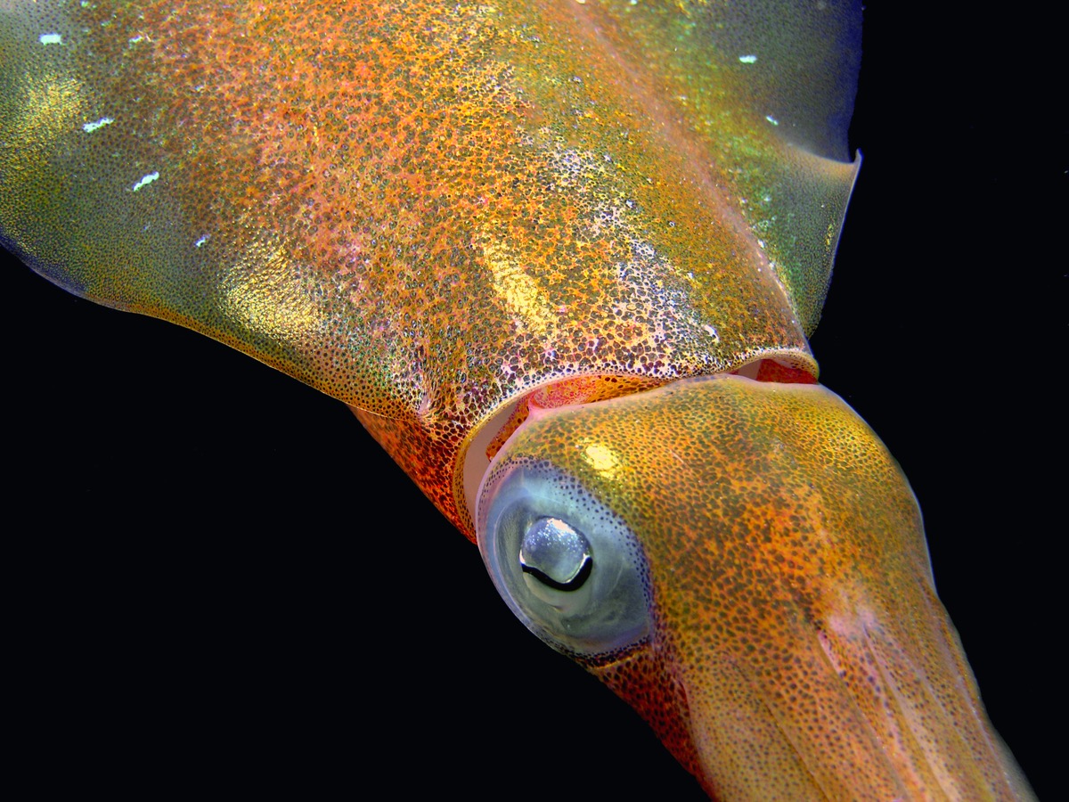
Figure 4. A fold built to move. A Caribbean reef squid at Bari Reef, Bonaire. The shimmer in its skin comes from light bouncing off stacked plates of folded protein, not from pigment. Photo by Betty Wills (2005), CC BY-SA 4.0, via Wikimedia Commons.

A fold can also be lost forever. An egg cracked into a hot pan turns from clear to white in seconds. Heat shakes the egg’s proteins out of their folds, and the open chains grab one another into a solid mesh. Cooled on the plate, the white never turns clear again. Anfinsen’s chains had a dilute bath and room to fold back. In the pan, each open chain meets its neighbors first. Not one letter in the egg changed. Some folds go wrong with every letter in place.
When a Fold Copies Itself
At a wedding banquet, the staff fold hundreds of napkins to match one sample on the first table. If the sample is folded wrong, every napkin copied from it comes out wrong, and the bride’s aunt spots each one. The cloth is the same. Only the fold spreads.
Narrator: A prion, a protein that passes on its wrong fold, works like that sample, with no staff needed. Every person makes a protein called PrP on the surface of nerve cells, folded mostly into coils. Its rogue form has the same chain of amino acids, letter for letter, folded instead into flat sheets. When a rogue copy touches a normal one, it refolds the normal one to match, and each new rogue copy refolds more. The rogue copies pile into clumps that kill nerve cells and leave tiny holes all through the brain (Figure 5). Like the road map’s wrong creases, a prion’s fold survives the ordinary cleaning that sterilizes surgical tools. A few patients caught a prion disease from instruments used on an earlier patient’s brain.
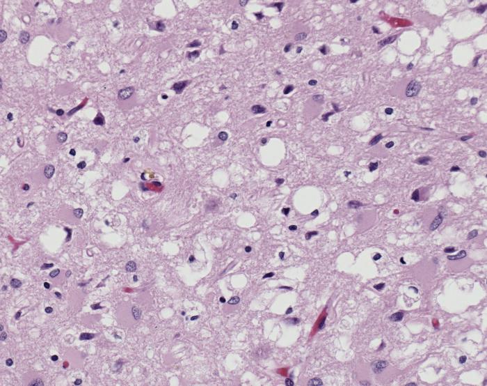
Figure 5. The holes a wrong fold leaves. Brain tissue from a case of vCJD, magnified 100 times. The white holes are the sponge-like change prions cause, and many nerve cells are gone. Photo by Sherif Zaki and Wun-Ju Shieh, CDC, public domain, via Wikimedia Commons.

In the 1950s, doctors in the highlands of Papua New Guinea met a disease the Fore people called kuru, from their word for shaking. Doctors began counting the dead in 1957. By the end of 1961, about a thousand people had died of it, most of them women and children. The Fore honored their dead by eating them at funeral feasts, and the women and children ate the brain. When the feasts stopped in the 1950s, kuru slowly died out. In 1966, Carleton Gajdusek’s team passed it to chimpanzees with brain tissue from patients. In 1982, Stanley Prusiner named the agent a prion, short for an infectious particle made of protein, with no genes of its own.
In 1986, British vets found a new brain disease in cattle, mad cow disease, spread through feed made from the remains of other cattle. In 1996, doctors reported ten young people in the UK with a new form of a human prion disease. It was named variant Creutzfeldt-Jakob disease (vCJD), and it most likely came from beef. In 1999, to keep vCJD out of the US blood supply, the FDA asked blood banks to turn away anyone with six months in the UK. In 2002 the line tightened to three months. Three people in the UK later caught vCJD from a blood transfusion, among the 178 who have died of it there. In 2022 the FDA lifted the deferral.
A prion passes on a fold, not a sequence: a shape copies itself. Other proteins fold wrong and stack too. In Alzheimer’s disease, a short protein called amyloid beta stacks its sheets into the sticky clumps that build up between nerve cells. In 2024, doctors in London described five people who developed Alzheimer’s decades after childhood treatment with growth hormone taken from the brains of the dead. Nothing suggests Alzheimer’s spreads between people in daily life. Can a drug stop a wrong fold, the way it stops a germ?
When the Builders Jam
A dose of azithromycin, a common antibiotic, reaches a germ and jams its builders. Bacteria have builders too, and many antibiotics work this way. The drug clips onto a bacterial ribosome and stops it from reading, and a bacterium that cannot build its proteins cannot grow. Human ribosomes are built differently enough that the drug leaves the body’s own mostly alone.
A cold comes from a virus, and a virus has no builders of its own. It hands its thread to the body’s builders. When a parent brings in a child with a sore throat, one question decides whether an antibiotic can help: is this infection bacterial? Viruses cause most sore throats, and most get better on their own within a week. Strep throat is the common exception, and a quick swab can tell. An antibiotic taken for a cold finds nothing to jam.
A prion gives a drug nothing to jam either: a prion has no builders, just a crease that spreads. Against a fold that copies itself, the guard a blood drive had was a question on a form.
Not every chain the builders make stays where it was built. One of them folds into an imp named Hush, who lays caps on the letters and decides which verses get read at all. A chain the builders let go comes back to choose which threads the builders will be handed next.
A Closing Invitation. Stanley stood for a protein: the same chain, in a new fold, does a new job, and a wrong fold can copy itself. One chain comes back as three checks: its builders, its copied fold, its letter.
- Look at your last antibiotic. Open the pharmacy app on your phone, or turn the bottle in the cabinet, and watch its date come up. Was it for a cold, a scratchy throat, a cough that cleared in a week? Ask the builders’ question: was that infection bacterial? What will you ask at the next cold?
- Take the chair back. If a blood drive at work or at church once turned you away from the chair for time in the UK, call the blood bank this month. Ask to be checked again. The question is gone. Who in your family heard that no, and would take the next chair beside you?
- Ask about the sticky letter. Before a pregnancy, yours or a sibling’s, ask at your next checkup whether either partner carries one copy of the sickle cell letter. A simple blood test, one pinch in the arm, finds it. Two carriers have a one-in-four chance with each child. Who in your family already knows their result?
Stanley went back to round with a pump, and a donor turned away can ask for the chair back with one call. Many proteins fold back on their own, unless a wrong fold has taught them another.
Most working parts of your body are proteins: chains folded into shape, the way creases make a paper airplane fly. The crease decides the job, and a wrong crease can spread to other chains of the same kind.
Where This Came From
In 1958, Francis Crick set down what he called the central dogma: “once ‘information’ has passed into protein it cannot get out again.” He meant one thing by information: “the precise determination of sequence.” Prusiner’s prions looked at first like a break in that rule, an infection with no genes. A prion copies no sequence, though. It hands its fold to a chain the cell built from its own gene, so Crick’s rule holds.
Intellectual Honesty Note. The sprites, imps, droplets and the field are invented staging; each mechanism they act out is standard biology, checked in the Narrator’s asides. The ribosome reads codons with the help of transfer RNAs, which the builders’ shelf stands in for. Anfinsen’s experiment used ribonuclease, and his 1961 work was summed up in his 1973 paper. Reflectins change color as many molecules shift together, not as one fold. How a rogue fold kills nerve cells is still debated. The prion form’s coil and sheet contents are from one 1993 measurement. The Flat Stanley plot follows summaries of the 1964 book.
References
AABB Donor History Task Force. (2005). Full-length donor history questionnaire (Version 1.1). AABB.
Alpers, M. P. (2008). The epidemiology of kuru: Monitoring the epidemic from its peak to its end. Philosophical Transactions of the Royal Society B, 363(1510), 3707–3713.
Anfinsen, C. B. (1973). Principles that govern the folding of protein chains. Science, 181(4096), 223–230.
Banerjee, G., Farmer, S. F., Hyare, H., Jaunmuktane, Z., Mead, S., Ryan, N. S., et al. (2024). Iatrogenic Alzheimer’s disease in recipients of cadaveric pituitary-derived growth hormone. Nature Medicine, 30(2), 394–402.
Brown, J. (1964). Flat Stanley (T. Ungerer, Illus.). Harper & Row.
Brown, P., Brandel, J.-P., Sato, T., Nakamura, Y., MacKenzie, J., Will, R. G., et al. (2012). Iatrogenic Creutzfeldt-Jakob disease, final assessment. Emerging Infectious Diseases, 18(6), 901–907.
Centers for Disease Control and Prevention. (n.d.). Healthy habits: Antibiotic do’s and don’ts. Retrieved October 5, 2026, from https://www.cdc.gov/antibiotic-use/about/index.html
Centers for Disease Control and Prevention. (2024, April 17). Sore throat basics. Retrieved October 17, 2026, from https://www.cdc.gov/sore-throat/about/index.html
Centers for Disease Control and Prevention. (2024, May 15). What is sickle cell trait? Retrieved October 17, 2026, from https://www.cdc.gov/sickle-cell/sickle-cell-trait/index.html
Crick, F. H. C. (1958). On protein synthesis. Symposia of the Society for Experimental Biology, 12, 138–163.
DeMartini, D. G., Izumi, M., Weaver, A. T., Pandolfi, E., & Morse, D. E. (2015). Structures, organization, and function of reflectin proteins in dynamically tunable reflective cells. Journal of Biological Chemistry, 290(24), 15238–15249.
Eaton, W. A., & Hofrichter, J. (1990). Sickle cell hemoglobin polymerization. Advances in Protein Chemistry, 40, 63–279.
Food and Drug Administration. (2022). Recommendations to reduce the possible risk of transmission of Creutzfeldt-Jakob disease and variant Creutzfeldt-Jakob disease by blood and blood components: Guidance for industry. Notice of availability, Federal Register, 87(100), May 24, 2022.
Gajdusek, D. C., Gibbs, C. J., & Alpers, M. (1966). Experimental transmission of a kuru-like syndrome to chimpanzees. Nature, 209(5025), 794–796.
Hartl, F. U., Bracher, A., & Hayer-Hartl, M. (2011). Molecular chaperones in protein folding and proteostasis. Nature, 475(7356), 324–332.
Ingram, V. M. (1957). Gene mutations in human haemoglobin: The chemical difference between normal and sickle cell haemoglobin. Nature, 180(4581), 326–328.
McGee, H. (2004). On food and cooking: The science and lore of the kitchen (Rev. ed.). Scribner.
National CJD Research & Surveillance Unit. (2025). Variant CJD cases worldwide and CJD monthly statistics. University of Edinburgh. Retrieved October 17, 2026, from https://www.cjd.ed.ac.uk/surveillance/data-and-reports
National Human Genome Research Institute. (n.d.). Codon. In Talking glossary of genomic and genetic terms. Retrieved October 17, 2026, from https://www.genome.gov/genetics-glossary/Codon
National Human Genome Research Institute. (n.d.). Translation. In Talking glossary of genomic and genetic terms. Retrieved October 4, 2026, from https://www.genome.gov/genetics-glossary/Translation
National Library of Medicine. (2000). Proteome. In Medical Subject Headings (MeSH, D020543). Retrieved October 17, 2026, from https://meshb.nlm.nih.gov/record/ui?ui=D020543
Nissen, P., Hansen, J., Ban, N., Moore, P. B., & Steitz, T. A. (2000). The structural basis of ribosome activity in peptide bond synthesis. Science, 289(5481), 920–930.
Pan, K.-M., Baldwin, M., Nguyen, J., Gasset, M., Serban, A., Groth, D., et al. (1993). Conversion of alpha-helices into beta-sheets features in the formation of the scrapie prion proteins. Proceedings of the National Academy of Sciences, 90(23), 10962–10966.
Pauling, L., & Corey, R. B. (1951). The pleated sheet, a new layer configuration of polypeptide chains. Proceedings of the National Academy of Sciences, 37(5), 251–256.
Pauling, L., Corey, R. B., & Branson, H. R. (1951). The structure of proteins: Two hydrogen-bonded helical configurations of the polypeptide chain. Proceedings of the National Academy of Sciences, 37(4), 205–211.
Prusiner, S. B. (1982). Novel proteinaceous infectious particles cause scrapie. Science, 216(4542), 136–144.
Wells, G. A. H., Scott, A. C., Johnson, C. T., Gunning, R. F., Hancock, R. D., Jeffrey, M., et al. (1987). A novel progressive spongiform encephalopathy in cattle. Veterinary Record, 121(18), 419–420.
Will, R. G., Ironside, J. W., Zeidler, M., Cousens, S. N., Estibeiro, K., Alperovitch, A., et al. (1996). A new variant of Creutzfeldt-Jakob disease in the UK. The Lancet, 347(9006), 921–925.
Wills, B. (2005). Caribbean Reef Squid colors [Photograph]. Wikimedia Commons. CC BY-SA 4.0.
Wilson, D. N. (2014). Ribosome-targeting antibiotics and mechanisms of bacterial resistance. Nature Reviews Microbiology, 12(1), 35–48.