On 7 October 1892, a chemist in Munich drank cholera in one gulp. Max von Pettenkofer was 74, with sugar in his urine and not one tooth left. His cup held a cubic centimeter of a fresh lab culture of comma-shaped bacteria. First, though, he drank a gram of baking soda stirred into Munich tap water, to blunt the stomach acid that kills the germ. The germ can drain a body of water through the gut within hours.
He meant to be the one who stayed well. In Berlin, Robert Koch had found the comma bacillus in cholera patients and said the germ was the cause. Pettenkofer thought a germ needed help: from the soil, the season and the person it entered. Within days he had colic and loose stools, and they lasted until 15 October. His stool was “swarming with comma bacilli.” Cholera itself never came, and he took that as proof.
He was wrong about the germ. Vibrio cholerae does cause cholera. His question was better than his answer: why does one person swallow a germ and stay well?
In 2018, Firas Midani, Regina LaRocque and colleagues asked it again in Dhaka, Bangladesh. They followed 76 people who lived under one roof with a cholera patient, for 30 days. Twenty-two caught the germ, and 54 did not. Swabs from each person’s gut, taken when they joined the study, told the two groups apart better than chance. The pattern lay in the crowd of gut bacteria, about 100 kinds. Those who got infected carried fewer of one large group, the Bacteroidetes. The swabs show who was more likely to catch the germ, not why.
In my house, no one is skipped. A cold comes home with my older daughter and goes through everyone, one at a time, until it has passed around the whole family. A cold is not cholera, and a house is not a study. Still, under my roof the germ wins every time, and in Dhaka most of the household never caught it.
Say an email goes round the office on a Monday: a stomach bug has hit the third floor. By Wednesday, half the desks on your row are empty. You ate the sandwiches at Monday’s meeting and filled your cup beside the people now home sick, and you stay well. You still cancel Saturday’s drive to your parents’, two hours each way, because you cannot tell why the bug passed you, or whether it will come back.
If the same germ reaches everyone under one roof and fewer than one in three catch it, what does the one who stays well carry that the others do not? The next cold that comes home is a chance to ask who it skips, and why.
Ziqi Ma and three colleagues gave one answer in 2024, in a review of the body’s microbes and human health. The first part of this series took two of their ideas: a person carries a second set of genes in their microbes, and a body without them has missing parts. Their next idea is about the host a germ meets, and that host is more than the person alone.
Who the Host Is
People who share a home also share some of their gut bacteria. With the bacteria comes part of the second set of genes, the adaptive genome. In 2023, Mireia Valles-Colomer and colleagues traced bacterial strains, the kinds within one species, across more than 9,700 samples from around the world. People in one home shared about 12 percent of their gut strains and 32 percent of their mouth strains. The longer they had lived together, the more mouth strains they shared.
That sharing begins on the day someone new moves under one roof: a new baby, a new partner, a new roommate. The question for a household is who brought what in, and the samples cannot answer it. They show who shares strains, not who passed them to whom.
In South Africa, Christina Gosmann and Douglas Kwon’s team tracked one germ, HIV, in 236 young women. They tested the women again and again. None had it at the start, and all were 18 to 23. Thirty-one became infected during the study.
The team had also sampled the bacteria in each woman’s vagina. Some women had a mixed community, with many kinds of bacteria and few Lactobacillus. Others had a community dominated by one species, Lactobacillus crispatus. The mixed group was more than four times as likely to become infected. Not one of the women dominated by L. crispatus became infected. The mixed communities came with more of the activated immune cells that HIV infects. A study that follows people cannot settle what caused what, though. The women with mixed communities may have differed in other ways too, and the bacteria could be a marker as much as a cause.
Animals can be tested more directly. In 1955, Bruce Phillips of the US Public Health Service and colleagues placed Entamoeba histolytica, an amoeba that causes dysentery, into the guts of germ-free guinea pigs. The amoeba did no harm. When the guinea pigs got one kind of common gut bacterium first, the amoeba damaged the gut wall.
Scott O’Neill’s team at Monash University ran a cleaner test, on a mosquito. Aedes aegypti carries dengue, a virus that brings fever, crushing joint pain and, in the worst cases, bleeding. Many insects carry a bacterium called Wolbachia inside their cells, but this mosquito normally does not. The team moved a strain of Wolbachia from fruit flies into Aedes aegypti. Inside the mosquitoes that carried it, the virus grew poorly. Like the aphid’s bacteria in part one, Wolbachia passes from mother to eggs, so it can spread through a wild population on its own.
From 2017 to 2020, Adi Utarini and colleagues at Gadjah Mada University tested those mosquitoes in Yogyakarta, Indonesia. They split a part of the city, home to about 313,000 people, into 24 areas and released Wolbachia mosquitoes in half of them, chosen at random. Volunteers left buckets of water and mosquito eggs around the neighborhoods, and the eggs hatched into mosquitoes carrying the bacterium. In the areas that got them, dengue fell by 77 percent, and hospital stays for dengue by 86 percent. It was one trial, in one city. With the same mosquito and the same virus, the bacteria living inside the mosquito changed how often it passed the virus on.
Whether a germ makes someone sick depends on the germ, on the person’s own genes and on the microbes already there. Ma calls the person and their microbes together a meta-host. Doctors have long asked why one germ makes one patient very sick and leaves another well, and why one transplant takes while another fails. Ma’s team points to the meta-host as part of the answer. Pettenkofer’s cup asked the same thing in 1892, and the Dhaka swabs asked it again in 2018. The germ was Koch’s. The host was Pettenkofer and everything living in his gut.
When a stomach bug goes through a home of four and skips one person, that person is a different meta-host from the other three. The crowd that person carries is one of three places to look for why. The host had always been a crowd, and part of that crowd lives in every human mouth.
What the Body Keeps in Check
On 17 September 1683, Antonie van Leeuwenhoek wrote to the Royal Society about the white paste on his teeth. He was a cloth merchant in Delft who ground his own lenses. He scraped some of the paste from between his teeth, thick as wet flour, and looked at it through one of his lenses. It was alive.
Some of the tiny animals darted through it “as a Jack or Pike does thro the water.” Others “spun about like a Top.” He kept his teeth “usually very clean,” he wrote, and still the animals were there. He looked at the teeth of two old men, one who lived soberly and one who drank brandy and wine every day, and found animals there too.
Three centuries later, scientists still argue about what to call them. In 1992, the immunologist Velio Bocci, at the University of Siena in Italy, called the gut’s bacteria “the neglected organ,” and the label spread. In 2022, Alessandro Fucarino, Francesco Cappello and colleagues at the University of Palermo answered that the label does not fit. An organ is built from tissues that grow from the embryo, and microbes arrive from outside. In a 2026 essay, the microbial ecologist Allan Konopka points out that the right picture depends on the level someone looks from.
In their 2024 review, Ma’s team looks from the level of tissues. Next to the four kinds in biology textbooks, epithelial (the body’s linings), connective, muscle and nervous, they propose a fifth. They call it a “slave tissue,” a term borrowed from sociology, to say the body is in charge. This piece calls it a managed tissue.
The managing never stops, because bacteria left alone keep multiplying. More cells make more cells, a reinforcing loop that slows only when the food runs out. So the body sets limits of its own, and one of them can be seen under a microscope.
In 2008, Malin Johansson, Gunnar Hansson and colleagues at the University of Gothenburg, in Sweden, looked at the lining of the colon in five mice. It wore two coats of mucus, together about as thick as two human hairs. The outer coat was loose and full of bacteria. The inner coat, packed tight and stuck to the wall, held none. In mice bred without the main mucus protein, bacteria lay against the cells of the wall, and the colon became inflamed.
Mucus is one limit among several. Antibodies released into the gut keep microbes in their places. Proteins in the gut and in milk lock away iron and zinc that bacteria need to grow. Stomach acid kills many germs before they reach the gut, which is why Pettenkofer drank his baking soda first. Leeuwenhoek’s tooth cleaning in Delft belongs on that list. So do the milk sugars that opened this series. Only some bacteria can eat them, so a mother’s milk helps choose which ones grow in her baby’s gut. What the one who stays well carries is a crowd, and the crowd helps only while the body keeps it in check.
The same managing reaches the dinner plate. Onions, apples and whole-grain bread carry fiber, food the body cannot digest and many gut bacteria can. When fiber runs short, some of those bacteria turn to the mucus coat instead (Figure 1). What goes on the plate at this week’s seven dinners helps decide what the colon’s bacteria eat.
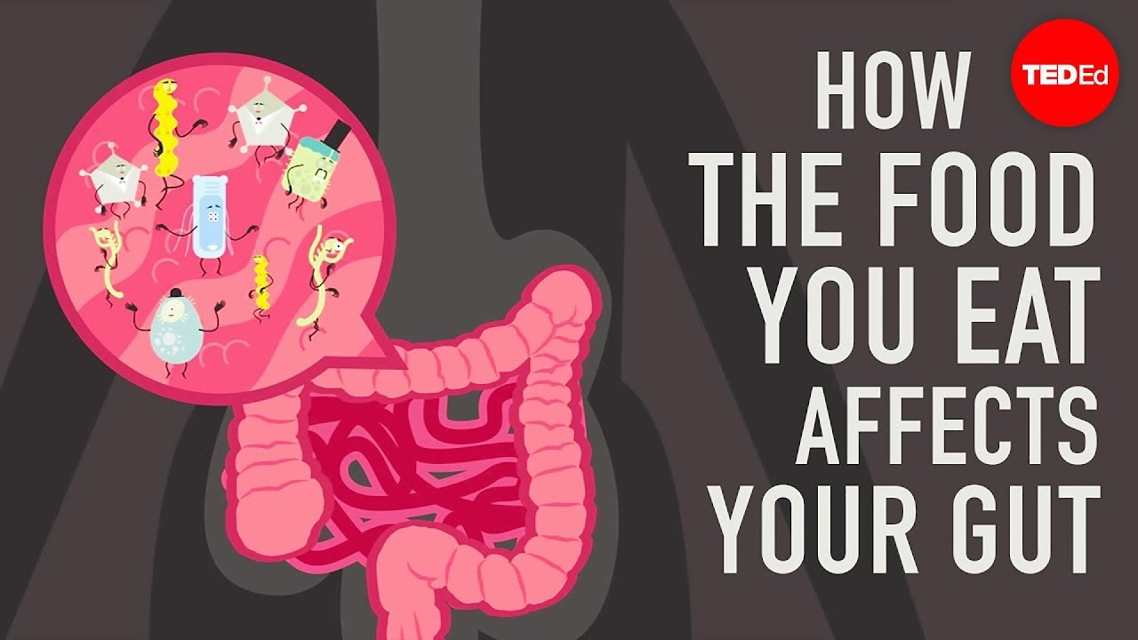
Figure 1. Fiber as food for gut bacteria, and what hungry bacteria do to the mucus lining, from TED-Ed (2017): lesson by Shilpa Ravella, animation by Andrew Foerster. CC BY-NC-ND 4.0.
By the sink, a toothbrush with splayed bristles has been doing the managing twice a day. No brush reaches the gut wall.
Where the Oxygen Goes
Right next to the gut wall, there is almost no oxygen, and the body’s own cells keep it that way. The cells that line the colon run mostly on butyrate, made by gut bacteria from fiber like the onion’s and the apple’s. Butyrate is the molecule that sealed a leaky brain barrier in part one. Its name comes from butter, and its acid gives rancid butter its sour smell.
Burning butyrate takes oxygen, and the cells take that oxygen from the edge of the gut. That suits the bacteria that make butyrate, because most of them cannot live where there is oxygen. The layer by the wall works like a shared room. The bacteria feed the cells, and the cells keep the room low on oxygen. The room runs on a reinforcing loop: each side keeps the other going.
In 2017, Mariana Byndloss, Andreas Bäumler and colleagues at the University of California, Davis, broke the loop with antibiotics in mice. The drugs killed the butyrate makers. Without butyrate, the colon cells switched to burning sugar, which uses less oxygen, and more oxygen reached the gut. E. coli and Salmonella, a germ behind food poisoning, can breathe that oxygen, and they multiplied. In Ma’s review, the life that gut cells and microbes share has a name: cell-microbe co-ecology. In that co-ecology, when one side shifts, the room shifts, and the room decides who can live in it. A germ that the room kept out yesterday can move in today. The one who stayed well last month can fall sick this month, with nothing changed in the germ.
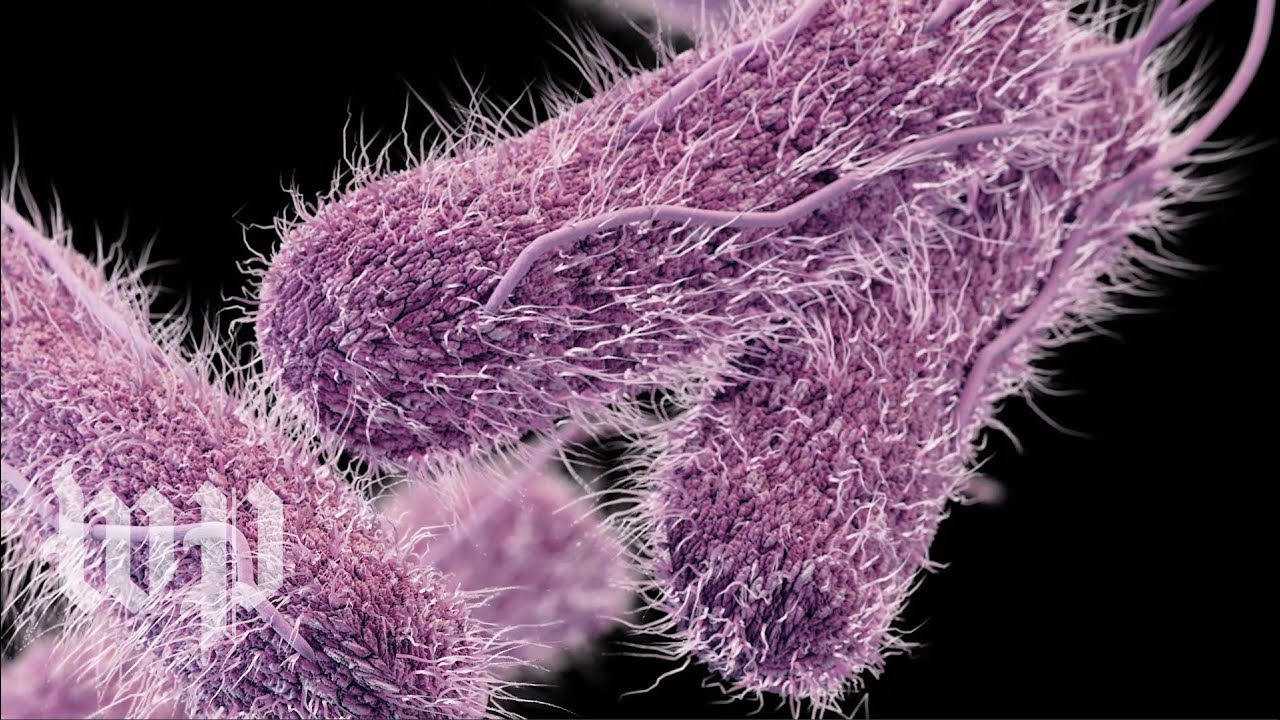
Figure 2. Salmonella, one of the germs that move in when oxygen reaches the gut, from The Washington Post (2018). Video and thumbnail: The Washington Post, Standard YouTube License.
The Davis work was done in mice, but an antibiotic course in a person can thin the butyrate makers too. The risk outlasts the pills. In the month after an antibiotic, a person is up to 10 times more likely to catch Clostridioides difficile, a gut germ that multiplies when antibiotics clear out its rivals. Age over 65 and a stay in a hospital or nursing home raise the risk again, and in older patients, C. difficile can be life-threatening.
In the days after the pills, loose stools are one sign the room has shifted. The CDC’s rule is short: diarrhea during an antibiotic course, or after it, is a reason to contact a doctor. A grandparent in a nursing home, just through a course, is the person most worth reminding of that rule. The germ may be the same as last month’s. The room it reaches has changed.
A Closing Reflection. Pettenkofer’s cup stood for an old question: why one germ takes one person and passes another. Part of the answer is the crowd each person carries, and the body that keeps it in check. Three ways to see yours.
- Map who shares. From where you sit, find one thing your home or office passes around: the mug by your hand, the toothbrush cup, the shared towel, the spoon at dinner. Who has lived or worked beside you longest, and what crowd do you two now carry in common?
- Trace the last bug. When the next cold reaches your home, think back, alone or with a sister or housemate, to the last stomach bug that went round. Who caught it first, who caught it last, and who was skipped? The same germ had its chance at each of you. What might the one who stayed well have been carrying?
- Feed the second crowd. At dinner tonight, slice an apple or an onion onto your plate and hear the knife crunch through. Which dish fed only you, and which fed your crowd too, so fewer of its bacteria turned to the mucus coat?
Tracing a bug through your house asks Pettenkofer’s question of everyone at once. When it has gone round, one bed still has its sheets on. The germ reached that bed too, and met a different crowd there.
Every germ arrives late. Each body’s gut bacteria got there first, so one stomach bug can go through a whole house and leave one person well.

Where This Came From
Of Ma’s eight ideas, this part takes three; the one on how resident microbes keep germs out and teach the immune system opens part three. Ma’s review gives cell-microbe co-ecology no section of its own; it gets one here. Pettenkofer’s side of the 1892 argument held that a germ needs the right soil, in the ground and in the person. The ground half did not hold up. The person half is the one this piece follows.
Intellectual Honesty Note. Pettenkofer’s dose, the baking soda, his symptoms and the “swarming” line come from the 1892 English report of his experiment, as quoted by Cabinet; the German original was not opened. “Stayed well” means he never had cholera; he had colic and loose stools for about a week. The office in the stakes paragraph is hypothetical. The Dhaka swabs were rectal swabs. The meta-host, the managed tissue and cell-microbe co-ecology are Ma’s proposals, not tested models. The mucus result is from five mice, not people. “Two human hairs” is this piece’s gloss of the mucus coats’ 150 micrometers. The Entamoeba result is reported from summaries of the paper, which was not opened. Leeuwenhoek’s words are from the Royal Society’s English abstract of his letter, printed in 1684.
References
Bocci, V. (1992). The neglected organ: Bacterial flora has a crucial immunostimulatory role. Perspectives in Biology and Medicine, 35(2), 251–260.
Burnett, D. G. (2009). Ingestion / The X factor. Cabinet, 33. https://cabinetmagazine.org/issues/33/burnett.php
Byndloss, M. X., Olsan, E. E., Rivera-Chávez, F., Tiffany, C. R., Cevallos, S. A., Lokken, K. L., Torres, T. P., Byndloss, A. J., Faber, F., Gao, Y., Litvak, Y., Lopez, C. A., Xu, G., Napoli, E., Giulivi, C., Tsolis, R. M., Revzin, A., Lebrilla, C. B., & Bäumler, A. J. (2017). Microbiota-activated PPAR-γ signaling inhibits dysbiotic Enterobacteriaceae expansion. Science, 357(6351), 570–575.
Centers for Disease Control and Prevention. (2026). About C. diff. https://www.cdc.gov/c-diff/about/index.html
Fucarino, A., Burgio, S., Paladino, L., Caruso Bavisotto, C., Pitruzzella, A., Bucchieri, F., & Cappello, F. (2022). The microbiota is not an organ: Introducing the muco-microbiotic layer as a novel morphofunctional entity. Anatomia, 1, 186–203.
Gosmann, C., Anahtar, M. N., Handley, S. A., Farcasanu, M., Abu-Ali, G., Bowman, B. A., et al. (2017). Lactobacillus-deficient cervicovaginal bacterial communities are associated with increased HIV acquisition in young South African women. Immunity, 46(1), 29–37.
Indriani, C., Tanamas, S. K., Khasanah, U., Ansari, M. R., Rubangi, Tantowijoyo, W., et al. (2023). Impact of randomised wMel Wolbachia deployments on notified dengue cases and insecticide fogging for dengue control in Yogyakarta City. Global Health Action, 16(1), 2166650. https://doi.org/10.1080/16549716.2023.2166650
Johansson, M. E. V., Phillipson, M., Petersson, J., Velcich, A., Holm, L., & Hansson, G. C. (2008). The inner of the two Muc2 mucin-dependent mucus layers in colon is devoid of bacteria. Proceedings of the National Academy of Sciences, 105(39), 15064–15069. https://doi.org/10.1073/pnas.0803124105
Konopka, A. (2026, May 6). Systems thinking [Substack post]. https://thinkmicrobe.substack.com/p/systems-thinking
Leeuwenhoek, A. van. (1684). An abstract of a letter from Mr. Anthony Leevvenhoeck at Delft, dated Sep. 17. 1683, containing some microscopical observations, about animals in the scurf of the teeth. Philosophical Transactions of the Royal Society of London, 14(159), 568–574. https://doi.org/10.1098/rstl.1684.0030
Ma, Z., Zuo, T., Frey, N., & Rangrez, A. Y. (2024). A systematic framework for understanding the microbiome in human health and disease: From basic principles to clinical translation. Signal Transduction and Targeted Therapy, 9, 237. https://doi.org/10.1038/s41392-024-01946-6
Midani, F. S., Weil, A. A., Chowdhury, F., Begum, Y. A., Khan, A. I., Debela, M. D., et al. (2018). Human gut microbiota predicts susceptibility to Vibrio cholerae infection. Journal of Infectious Diseases, 218(4), 645–653. https://doi.org/10.1093/infdis/jiy192
Phillips, B. P., Wolfe, P. A., Rees, C. W., Gordon, H. A., Wright, W. H., & Reyniers, J. A. (1955). Studies on the ameba-bacteria relationship in amebiasis: Comparative results of the intracecal inoculation of germfree, monocontaminated, and conventional guinea pigs with Entamoeba histolytica. American Journal of Tropical Medicine and Hygiene, 4(4), 675–692.
Ravella, S. (2017, March 23). How the food you eat affects your gut [Video]. TED-Ed. YouTube. https://www.youtube.com/watch?v=1sISguPDlhY
Utarini, A., Indriani, C., Ahmad, R. A., Tantowijoyo, W., Arguni, E., Ansari, M. R., et al. (2021). Efficacy of Wolbachia-infected mosquito deployments for the control of dengue. New England Journal of Medicine, 384(23), 2177–2186.
Valles-Colomer, M., Blanco-Míguez, A., Manghi, P., Asnicar, F., Dubois, L., Golzato, D., et al. (2023). The person-to-person transmission landscape of the gut and oral microbiomes. Nature, 614(7946), 125–135.
Walker, T., Johnson, P. H., Moreira, L. A., Iturbe-Ormaetxe, I., Frentiu, F. D., McMeniman, C. J., et al. (2011). The wMel Wolbachia strain blocks dengue and invades caged Aedes aegypti populations. Nature, 476(7361), 450–453.
The Washington Post. (2018, August 30). What is salmonella? [Video]. YouTube. https://www.youtube.com/watch?v=WTlFIwQePkg